Cardiovascular System for TEAS 7: Blood Flow Pathway & Heart Anatomy

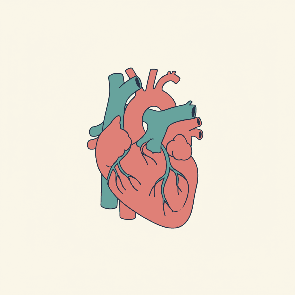
The Highest-Yield System on the TEAS
If you learn one body system thoroughly for the TEAS 7, make it the cardiovascular system. Tutors who track thousands of student results report that heart, vessel, and blood-flow questions appear on virtually every exam — typically 3 to 5 questions in the Science section — and the blood-flow pathway is the single most repeated question pattern in all of Anatomy and Physiology. It also underpins your future nursing work, from reading blood pressures to understanding cardiac medications.
This guide gives you the complete system the way the TEAS tests it: heart chambers, the four valves, the 14-step blood-flow pathway with a memory trick, the artery-versus-vein exception designed to trap you, blood pressure, and the heart's electrical conduction system — plus worked examples in true exam style.
Heart Chambers: Receive on Top, Pump Below
The heart is a four-chambered muscular pump about the size of your fist. One rule anchors everything: atria receive, ventricles pump. The thin-walled upper atria collect returning blood; the muscular lower ventricles eject it.
| Chamber | Receives from | Pumps to | Blood type |
|---|---|---|---|
| Right atrium | Body via vena cava | Right ventricle | Deoxygenated |
| Right ventricle | Right atrium | Lungs via pulmonary artery | Deoxygenated |
| Left atrium | Lungs via pulmonary veins | Left ventricle | Oxygenated |
| Left ventricle | Left atrium | Body via aorta | Oxygenated |
The left ventricle has the thickest muscular wall of all four chambers because it must generate enough force to push blood through the entire body. When a question asks which chamber is strongest, the answer is always the left ventricle.
The Four Valves: One-Way Doors
Valves keep blood moving forward and prevent backflow. Two atrioventricular valves sit between atria and ventricles; two semilunar valves guard the exits into the great arteries.
| Valve | Type | Location |
|---|---|---|
| Tricuspid | Atrioventricular | Right atrium to right ventricle |
| Pulmonary | Semilunar | Right ventricle to pulmonary artery |
| Mitral (bicuspid) | Atrioventricular | Left atrium to left ventricle |
| Aortic | Semilunar | Left ventricle to aorta |
Memorize the valve order with the mnemonic "Try Pulling My Aorta" — Tricuspid, Pulmonary, Mitral, Aortic. Write it on your scratch paper the moment the exam starts; sequence questions then become simple lookups.
The 14-Step Blood Flow Pathway
Tracing a drop of blood through the heart is the most tested cardiovascular skill. Walk it deliberately until the order is automatic:
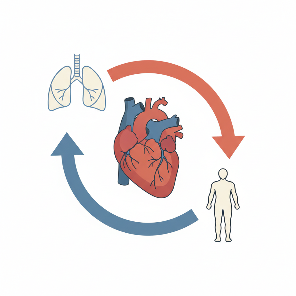
- Deoxygenated blood enters the right atrium via the superior and inferior vena cava.
- It passes the tricuspid valve into the right ventricle.
- The right ventricle pumps it through the pulmonary valve into the pulmonary arteries.
- Blood reaches the lungs, releases carbon dioxide, and picks up oxygen.
- Oxygenated blood returns through the pulmonary veins to the left atrium.
- It passes the mitral (bicuspid) valve into the left ventricle.
- The left ventricle pumps it through the aortic valve into the aorta.
- The aorta distributes oxygen-rich blood to the entire body.
The heart runs two loops at once. The pulmonary circuit (heart to lungs and back) handles gas exchange; the systemic circuit (heart to body and back) delivers oxygen and collects waste. Blood passes through the heart twice per full lap, which is why circulation is called a double loop.
Arteries vs. Veins: The Exception That Traps Students
Forget the shortcut that arteries carry oxygenated blood. The TEAS-safe rule is directional: arteries carry blood away from the heart; veins carry blood toward it.
| Vessel | Direction | Walls | Usual blood |
|---|---|---|---|
| Arteries | Away from heart | Thick, elastic, high pressure | Oxygenated — except pulmonary |
| Veins | Toward heart | Thinner, with valves | Deoxygenated — except pulmonary |
| Capillaries | Connect both | One cell thick | Exchange site for gases and nutrients |
The pulmonary vessels break the usual pattern: pulmonary arteries carry deoxygenated blood to the lungs, and pulmonary veins carry oxygenated blood back. Test writers love this exception — when a question describes a vessel carrying deoxygenated blood away from the heart, the answer is the pulmonary artery.
Blood Pressure and the Heart's Electrical System
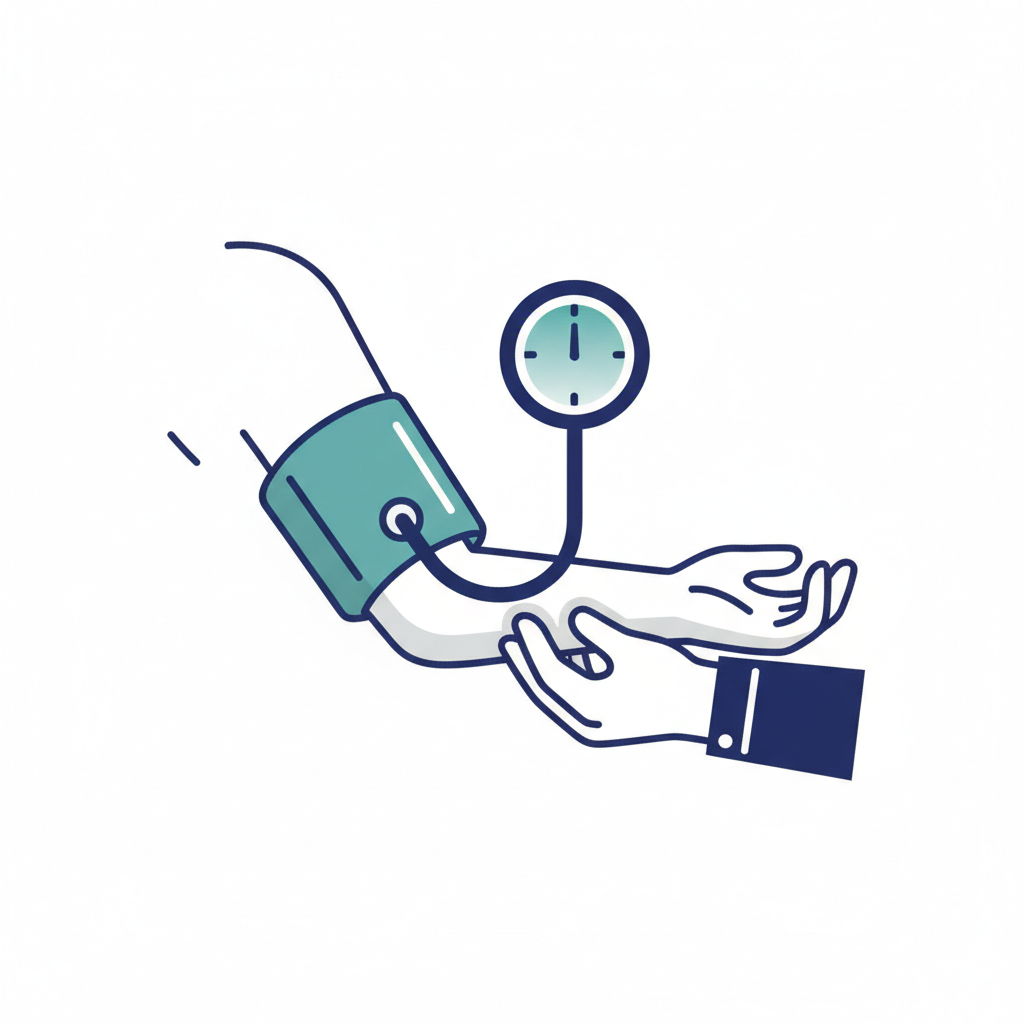
Blood pressure is the force of blood against arterial walls, written systolic over diastolic (for example, 120/80 mmHg). The systolic top number measures pressure during ventricular contraction; the diastolic bottom number measures pressure during relaxation. Normal resting values sit near 120/80.
Each beat is triggered by an electrical conduction sequence worth memorizing in order: SA node (the natural pacemaker, firing 60–100 times per minute) → AV node → Bundle of His → bundle branches → Purkinje fibers, which spread the impulse through the ventricles. Also know the cardiac output formula: cardiac output = heart rate × stroke volume, roughly 5 liters per minute at rest.
Worked Examples in Exam Style
Example 1: Blood in the right ventricle will next flow to the — pulmonary artery. Locate yourself on the pathway: right ventricle pumps through the pulmonary valve into the pulmonary artery toward the lungs. Note the trap: that artery carries deoxygenated blood.
Example 2: Which chamber has the thickest wall? The left ventricle, because it pumps blood to the entire systemic circuit.
Example 3: A resting heart rate of 70 beats per minute with a stroke volume of 70 mL per beat gives a cardiac output of 70 × 70 = 4,900 mL per minute, or about 5 L/min — the normal resting value.
Study Checklist
- Recite the 14-step pathway from memory, forward and from any midpoint ("what comes after the mitral valve?").
- Know all four valves by name, type, and location — use "Try Pulling My Aorta."
- State the pulmonary exception in one sentence without hesitating.
- Recall the conduction order: SA node, AV node, Bundle of His, bundle branches, Purkinje fibers.
- Calculate cardiac output from heart rate and stroke volume.
Cardiovascular questions reward students who map the system rather than memorize isolated facts. Draw the pathway from memory once a day until it takes under a minute — that single exercise converts the most heavily tested TEAS topic from a risk into guaranteed points. For the exam-format side of anatomy — hotspot diagrams and ordered-sequence items — see our question types guide.
Frequently Asked Questions
Related Posts
View all articles
10 TEAS Mistakes That Cost Points (and How to Avoid Every One)
The 10 TEAS mistakes behind most lost points: planning, study strategy, execution, and test-day errors, plus an anti-mistake checklist.

Beat TEAS Test Anxiety: A Per-Section Pacing Plan That Works
Beat TEAS test anxiety with exact per-section pacing budgets, panic-proof preparation habits, and a 30-second mid-exam recovery protocol.

TEAS for LPN Programs: Scores, Paths & a 4-Week Plan
TEAS for LPN programs: realistic score targets, LPN vs RN paths, and a focused 4-week study plan for working adults.